MENU
TW | TWD
TW | TWD
You are about to leave this site.
Please be aware that your current cart is not saved yet and cannot be restored on the new site nor when you come back. If you want to save your cart please login in into your account.
No results found
Search Suggestions

How to identify Mycoplasma contamination in your cell culture
Lab Academy
- Cell Biology
- Cell Culture
- Contamination
- CO2 Incubators
- Cell Culture Consumables
- Essay
Macroscopic detection
Mycoplasma-positive cell cultures show no visible changes to the media.Microscopic detection
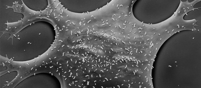
Mycoplasma are only about 0.1 - 0.3 µm in diameter, therefore detection via brightfield microscopy is not possible. This lack of visible signs of infection increases the risk of mycoplasma-positive cells remaining unnoticed.Experiments carried out with mycoplasma-infected cells may yield false, misleading and non-reproducible results. It is therefore crucial to test all cultures for mycoplasma on a regular basis. One simple method employs DNA staining; however, this method presents certain drawbacks. This table provides an overview of the advantages and disadvantages of different mycoplasma detection methods.
The method you will select may depend on one or more of the following:
- Access to the required equipment (for example thermocycler, fluorescence microscope, etc.).
- How many samples need to be tested at once.
- How urgently you need the results (the longer the testing procedure the higher the risk of spreading the contamination).

