菜单
CN | CNY
CN | CNY

How to identify fungal contamination in your cell culture
实验室学院
- 细胞生物学
- 细胞培养
- 污染
- 二氧化碳培养箱
- 细胞培养耗材
- 文章
Macroscopic detection
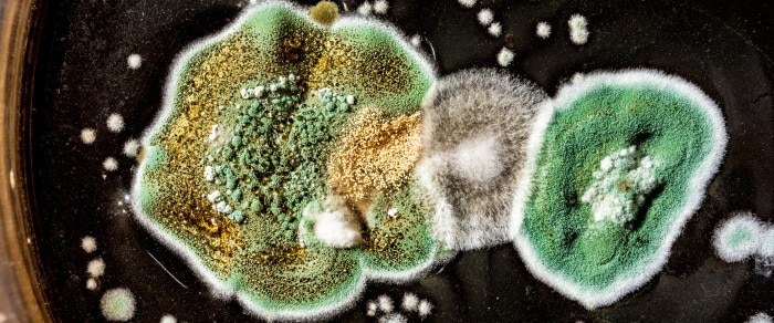
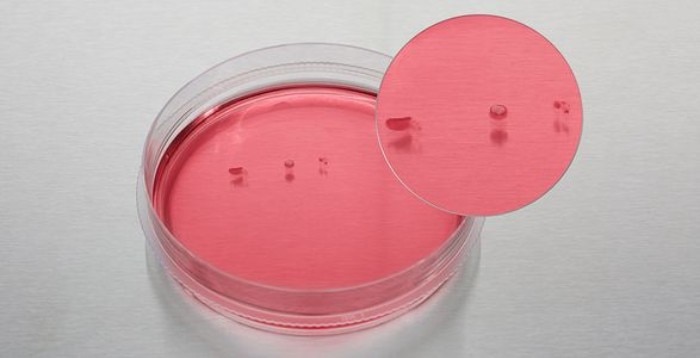
Fungi and mold can appear as small isolated colonies of grey, white or greenish color, floating at the surface of the medium. It is important to inspect the culture vessel with the naked eye and look for colonies which rest on the medium surface and will be missed during microscopic inspection. If the contamination is substantial, the medium will become turbid and cloudy, and spots on the vessel surface may appear. Sometimes fungal contaminations will cause a pH increase of the medium, resulting in phenol-red containing media to appear pink.
显示更少

Fungal colonies floating on the medium surface.
Microscopic detection
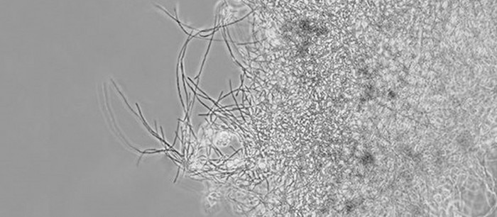
It is usually the fungal spores which get into a culture vessel and then begin to sprout and form the typical fiber like hyphae. Fungi growing on the bottom of the dish/flask are more easily detected in the microscope than those floating on the surface.
显示更少


