MENU
CA | CAD
-
-
-
- Services pour bioprocédés
- Services pour centrifugeuse et rotors
- Services pour Mastercycler
- Services pour automates de pipetage
- Services pour congélateurs
- Services pour incubateurs
- Services pour agitateurs
- Services pour appareils de photométrie
- Service de contrôle de la température et de l’agitation
- Service pour pipette
-
-
-
-
- Services pour bioprocédés
- Services pour centrifugeuse et rotors
- Services pour Mastercycler
- Services pour automates de pipetage
- Services pour congélateurs
- Services pour incubateurs
- Services pour agitateurs
- Services pour appareils de photométrie
- Service de contrôle de la température et de l’agitation
- Service pour pipette
-
CA | CAD
-
- Centrifugeuses de paillasse
- Centrifugeuses au sol
- Centrifugeuses réfrigérées
- Microcentrifugeuses
- Centrifugeuses multi-fonctions
- Centrifugeuses haute vitesse
- Ultracentrifugeuses
- Concentrateur
- High-Speed and Ultracentrifuge Consumables
- Tubes de centrifugeuse
- Plaques de centrifugeuse
- Gestion des appareils
- Gestion des échantillons et des informations
Aucun résultat trouvé
Chercher des suggestions

How to identify fungal contamination in your cell culture
Lab Academy
- Biologie cellulaire
- Culture cellulaire
- Contamination
- Incubateurs à CO2
- Consommables pour culture cellulaire
- Test
Macroscopic detection
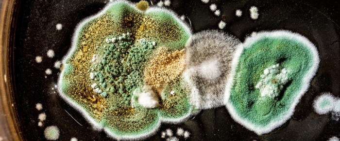
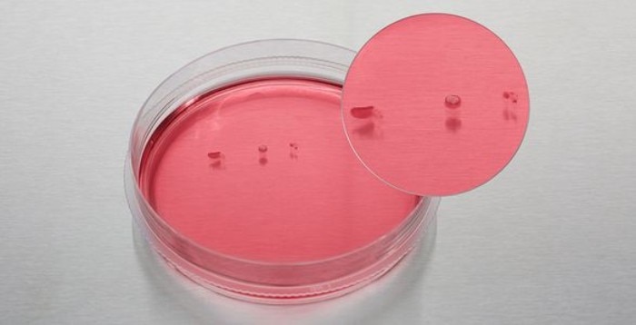
Fungi and mold can appear as small isolated colonies of grey, white or greenish color, floating at the surface of the medium. It is important to inspect the culture vessel with the naked eye and look for colonies which rest on the medium surface and will be missed during microscopic inspection. If the contamination is substantial, the medium will become turbid and cloudy, and spots on the vessel surface may appear. Sometimes fungal contaminations will cause a pH increase of the medium, resulting in phenol-red containing media to appear pink.
Lire moins

Fungal colonies floating on the medium surface.
Microscopic detection
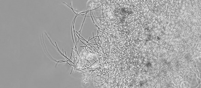
It is usually the fungal spores which get into a culture vessel and then begin to sprout and form the typical fiber like hyphae. Fungi growing on the bottom of the dish/flask are more easily detected in the microscope than those floating on the surface.
Lire moins

Related links
Lire moins

